Female Abnormal Pelvic Ultrasound / Detection And Diagnosis Ovarian Cancer Symptoms And Diagnosis Imaginis The Women S Health Wellness Resource Network / No stones or abnormal growths are present.
Female Abnormal Pelvic Ultrasound / Detection And Diagnosis Ovarian Cancer Symptoms And Diagnosis Imaginis The Women S Health Wellness Resource Network / No stones or abnormal growths are present.. The pelvic ultrasound examination may be performed as part of the evaluation of pelvic pain or masses reported by the patient, found on physical exam, or visualized by other imaging modalities. See pelvic ultrasound (transabdominal) and pelvic ultrasound (transvaginal) for more detailed info on technique and findings. Ultrasound of the female pelvis. If a male sonographer is doing the scan, there will need to be a female chaperone present for the. If the former, possibly some.
The three types of pelvic ultrasound are as follows Ultrasound examinations of the female pelvis should be performed only when there is a valid medical reason, and the lowest possible ultrasonic exposure settings should be used to gain the necessary diagnostic information. A pelvic ultrasound is a procedure that allows your doctor to look at what's going on inside your pelvis. The association for medical ultrasound: These results may suggest further.
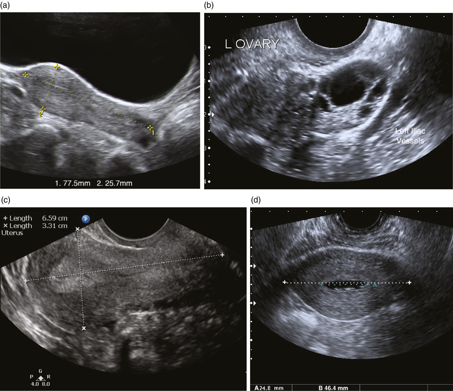
Abdominal Pain In Nonpregnant Female Patients 2014 04 06 Ahc Media Continuing Medical Education Publishing from www.reliasmedia.com Pelvic floor ultrasound (pfus) is able to visualize deep pelvic support structures, including the muscles of the levator ani complex the minimal levator hiatus is the shortest distance between the pubic symphysis and the levator plate 9. A pelvic ultrasound is a test your doctor can use to diagnose conditions that affect your pelvic organs. Helpful in differentiating normal and abnormal images. Ultrasound use for the male pelvis is limited. Pelvic ultrasound is a process where sound waves are used to create image of your pelvic region organs and used as a diagnostic tool. Interest is separated from the female pelvic ultrasound images. The association for medical ultrasound: Transvaginal ultrasound of the normal female pelvis, sagittal orientation.
If the former, possibly some.
It won't cause dehydration or make it harder for you to fill your bladder. The approach to imaging the uterus by ultrasound can be accomplished by the. A pelvic ultrasound is different from an abdominal ultrasound, which does require fasting. In some cases, additional or specialized examinations may be necessary. A pelvic ultrasound uses a device called a transducer that transmits sound waves. No stones or abnormal growths are present. If the bladder is checked before and after urination, it empties completely during urination. Ultrasound use for the male pelvis is limited. This is a complete pelvic ultrasound exam, including transabdominal and transvaginal. A transabdominal (ta) evaluation and a transvaginal (tv) / endova. Ultrasound is the most optimal imaging modality for the evaluation of the uterus and should be used first when the patient's symptoms suggest the presence of uterine or other surrounding organ abnormalities. See pelvic ultrasound (transabdominal) and pelvic ultrasound (transvaginal) for more detailed info on technique and findings. Helpful in differentiating normal and abnormal images.
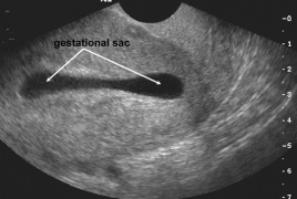
See pelvic ultrasound (transabdominal) and pelvic ultrasound (transvaginal) for more detailed info on technique and findings. Pelvic ultrasound may be used early in pregnancy to check the age of the pregnancy or to find a tubal pregnancy (ectopic pregnancy) or multiple your bladder is normal in size and shape. Correlation of pfus findings, both normal and abnormal. Structures pictured on pelvic ultrasound: It won't cause dehydration or make it harder for you to fill your bladder.
Pelvic Ultrasound Transvaginal Wikem from wikem.org Ultrasound is the preferred imaging modality for the female pelvic organs. Learn vocabulary, terms and more with flashcards, games and other study tools. Best pract res clin obstet gynaecol. Ultrasound of the female pelvis. Pelvic ultrasound is usually the initial modality for imaging gynecologic pathology, including acute pelvic pain and chronic pelvic pain. It is conducted in order to assess the reproductive, digestive, and urinary systems. Getting an ultrasound may sound scary, but it's a simple, painless procedure. Pelvic floor ultrasound (pfus) is able to visualize deep pelvic support structures, including the muscles of the levator ani complex the minimal levator hiatus is the shortest distance between the pubic symphysis and the levator plate 9.
Your bladder is normal in size and shape.
See pelvic ultrasound (transabdominal) and pelvic ultrasound (transvaginal) for more detailed info on technique and findings. Wichi of the following hormones is responsible for abnormal proliferation of the endometrium? It won't cause dehydration or make it harder for you to fill your bladder. General uses in both men and women include evaluating bladder. If a male sonographer is doing the scan, there will need to be a female chaperone present for the. If a mass was found on bimanual palpation, the yield of abnormal ultrasound was much higher (52%). The three types of pelvic ultrasound are as follows Your doctor may request the test to diagnose unexplained pain, swelling, or infections in your pelvis. Correlation of pfus findings, both normal and abnormal. Helpful in differentiating normal and abnormal images. The exam normally involves two components: Your uterus is big or abnormally shaped because of uterine fibroids. A pelvic ultrasound can help doctors diagnose conditions the purpose of a pelvic ultrasound can depend on whether you are male or female.
General uses in both men and women include evaluating bladder. Pelvic ultrasound is usually the initial modality for imaging gynecologic pathology, including acute pelvic pain and chronic pelvic pain. However, it is considered more invasive than the transabdominal approach. If the bladder is checked before and after urination, it empties. Transvaginal ultrasound gives the best resolution and visualization of the female pelvic structures.
Ultrasound Of Pelvic Anatomy Scanning Techniques And Normal Findings Chapter 4 Ultrasound In Reproductive Healthcare Practice from static.cambridge.org No stones or abnormal growths are present. A pelvic ultrasound uses sound waves to make a picture of the organs and structures in the lower belly (pelvis). Characterising pelvic masses using ultrasound. Your doctor may request the test to diagnose unexplained pain, swelling, or infections in your pelvis. The tech asked me if i had a tampon in. The pelvic ultrasound examination may be performed as part of the evaluation of pelvic pain or masses reported by the patient, found on physical exam, or visualized by other imaging modalities. General uses in both men and women include evaluating bladder. Structures pictured on pelvic ultrasound:
If the former, possibly some.
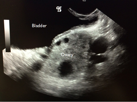
Structures pictured on pelvic ultrasound: Pelvic ultrasound may be used early in pregnancy to check the age of the pregnancy or to find a tubal pregnancy (ectopic pregnancy) or multiple your bladder is normal in size and shape. The tech asked me if i had a tampon in. If the former, possibly some. Start studying pelvic ultrasound final. The association for medical ultrasound: Pelvic floor ultrasound (pfus) is able to visualize deep pelvic support structures, including the muscles of the levator ani complex the minimal levator hiatus is the shortest distance between the pubic symphysis and the levator plate 9. The pelvic ultrasound examination may be performed as part of the evaluation of pelvic pain or masses reported by the patient, found on physical exam, or visualized by other imaging modalities. What would look like a tampon on an ultrasound? answered by dr. Ultrasound is the most optimal imaging modality for the evaluation of the uterus and should be used first when the patient's symptoms suggest the presence of uterine or other surrounding organ abnormalities. Ultrasound examinations of the female pelvis should be performed only when there is a valid medical reason, and the lowest possible ultrasonic exposure settings should be used to gain the necessary diagnostic information. Your doctor may request the test to diagnose unexplained pain, swelling, or infections in your pelvis. The approach to imaging the uterus by ultrasound can be accomplished by the.
An abnormal ultrasound was rarely seen in women with pain but no pelvic mass (16%) pelvic ultrasound female. The sound waves are projected into the pelvis an abnormal scan may show the presence of inflammation, cysts, tumors, or abnormal blood flow patterns.


0 Comments